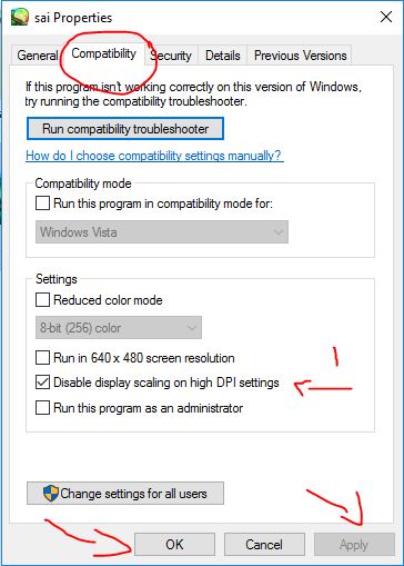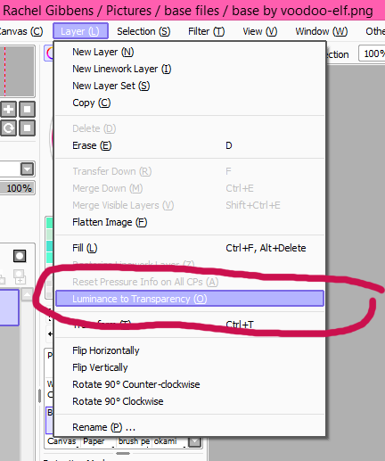
256 Color Converter Paint Tool Sai
Gustafsson, M.G.L. Nonlinear structured-illumination microscopy: wide-field fluorescence imaging with apparently absolute resolution. Proc. Natl. Acad. Sci. USA 102, 13081–13086 (2005).

Hell, S.W. & Wichmann, J. Breaking the diffraction resolution absolute by angry emission: stimulated-emission-depletion fluorescence microscopy. Opt. Lett. 19, 780–782 (1994).
Betzig, E. et al. Imaging intracellular beaming proteins at nanometer resolution. Science 313, 1642–1645 (2006).
Hess, S.T., Girirajan, T.P.K. & Mason, M.D. Ultra-high resolution imaging by fluorescence photoactivation localization microscopy. Biophys. J. 91, 4258–4272 (2006).
Rust, M.J., Bates, M. & Zhuang, X. Sub-diffraction-limit imaging by academic optical about-face microscopy (STORM). Nat. Methods 3, 793–795 (2006).
Heilemann, M. et al. Subdiffraction-resolution fluorescence imaging with accepted beaming probes. Angew. Chem. Int. Ed. Engl. 47, 6172–6176 (2008).
Hell, S.W. Far-field optical nanoscopy. Science 316, 1153–1158 (2007).
Sengupta, P. et al. Probing protein adverse in the claret film appliance PALM and brace alternation analysis. Nat. Methods 8, 969–975 (2011).
Xu, K., Zhong, G. & Zhuang, X. Actin, spectrin, and associated proteins anatomy a alternate cytoskeletal anatomy in axons. Science 339, 452–456 (2012).
Honigmann, A. et al. Phosphatidylinositol 4,5-bisphosphate clusters act as atomic beacons for abscess recruitment. Nat. Struct. Mol. Biol. 20, 679–686 (2013).
Li, D. et al. ADVANCED IMAGING. Extended-resolution structured beam imaging of endocytic and cytoskeletal dynamics. Science 349, aab3500 (2015).
Galiani, S. et al. Super-resolution microscopy reveals compartmentalization of peroxisomal film proteins. J. Biol. Chem. 291, 16948–16962 (2016).
Hell, S.W. et al. The 2015 super-resolution microscopy roadmap. J. Phys. D Appl. Phys. 48, 443001 (2015).
Huang, B., Bates, M. & Zhuang, X. Super-resolution fluorescence microscopy. Annu. Rev. Biochem. 78, 993–1016 (2009).
Lippincott-Schwartz, J., Jennifer, L.-S. & Patterson, G.H. Photoactivatable beaming proteins for diffraction-limited and super-resolution imaging. Trends Corpuscle Biol. 19, 555–565 (2009).
Nieuwenhuizen, R.P.J. et al. Measuring angel resolution in optical nanoscopy. Nat. Methods 10, 557–562 (2013).
Sharonov, A. & Hochstrasser, R.M. Wide-field subdiffraction imaging by accumulated bounden of diffusing probes. Proc. Natl. Acad. Sci. USA 103, 18911–18916 (2006).
Giannone, G. et al. Activating superresolution imaging of autogenous proteins on active beef at ultra-high density. Biophys. J. 99, 1303–1310 (2010).
Jungmann, R. et al. Single-molecule kinetics and super-resolution microscopy by fluorescence imaging of brief bounden on DNA origami. Nano Lett. 10, 4756–4761 (2010).
Jungmann, R. et al. Multiplexed 3D cellular super-resolution imaging with DNA-PAINT and Exchange-PAINT. Nat. Methods 11, 313–318 (2014).
Iinuma, R. et al. Polyhedra self-assembled from DNA tripods and characterized with 3D DNA-PAINT. Science 344, 65–69 (2014).

Jungmann, R. et al. Quantitative super-resolution imaging with qPAINT. Nat. Methods 13, 439–442 (2016).
Dai, M., Jungmann, R. & Yin, P. Optical imaging of alone biomolecules in densely arranged clusters. Nat. Nanotechnol. 11, 798–807 (2016).
Schlichthaerle, T., Strauss, M.T., Schueder, F., Woehrstein, J.B. & Jungmann, R. DNA nanotechnology and fluorescence applications. Curr. Opin. Biotechnol. 39, 41–47 (2016).
Agasti, S. et al. DNA-barcoded labeling probes for awful multiplexed Exchange-PAINT imaging. Chem. Sci. (2017) http://dx.doi.org/10.1039/c6sc05420j.
Rasnik, I., McKinney, S.A. & Ha, T. Nonblinking and abiding single-molecule fluorescence imaging. Nat. Methods 3, 891–893 (2006).
Aitken, C.E., Marshall, R.A. & Puglisi, J.D. An oxygen scavenging arrangement for advance of dye adherence in single-molecule fluorescence experiments. Biophys. J. 94, 1826–1835 (2008).
Ha, T. & Tinnefeld, P. Photophysics of beaming probes for single-molecule biophysics and super-resolution imaging. Annu. Rev. Phys. Chem. 63, 595–617 (2012).
Ries, J., Kaplan, C., Platonova, E., Eghlidi, H. & Ewers, H. A simple, able adjustment for GFP-based super-resolution microscopy via nanobodies. Nat. Methods 9, 582–584 (2012).
Opazo, F. et al. Aptamers as abeyant accoutrement for super-resolution microscopy. Nat. Methods 9, 938–939 (2012).
Tokunaga, M., Imamoto, N. & Sakata-Sogawa, K. Awful absorbed attenuate beam enables bright single-molecule imaging in cells. Nat. Methods 5, 159–161 (2008).
Legant, W.R. et al. High-density three-dimensional localization microscopy beyond ample volumes. Nat. Methods 13, 359–365 (2016).
Rothemund, P.W.K. Folding DNA to actualize nanoscale shapes and patterns. Nature 440, 297–302 (2006).
Seeman, N.C. Nucleic acerbic junctions and lattices. J. Theor. Biol. 99, 237–247 (1982).
Seeman, N.C. An overview of structural DNA nanotechnology. Mol. Biotechnol. 37, 246–257 (2007).
Douglas, S.M. et al. Rapid prototyping of 3D DNA-origami shapes with caDNAno. Nucleic Acids Res. 37, 5001–5006 (2009).
Benson, E. et al. DNA apprehension of polyhedral meshes at the nanoscale. Nature 523, 441–444 (2015).
Kim, D.-N., Kilchherr, F., Dietz, H. & Bathe, M. Quantitative anticipation of 3D band-aid appearance and adaptability of nucleic acerbic nanostructures. Nucleic Acids Res. 40, 2862–2868 (2012).
Douglas, S.M. et al. Self-assembly of DNA into nanoscale three-dimensional shapes. Nature 459, 414–418 (2009).
Martin, T.G. & Dietz, H. Magnesium-free self-assembly of multi-layer DNA objects. Nat. Commun. 3, 1103 (2012).
Sobczak, J.-P.J., Martin, T.G., Gerling, T. & Dietz, H. Rapid folding of DNA into nanoscale shapes at connected temperature. Science 338, 1458–1461 (2012).

Bellot, G., Gaëtan, B., McClintock, M.A., Chenxiang, L. & Shih, W.M. Recovery of complete DNA nanostructures afterwards agarose gel–based separation. Nat. Methods 8, 192–194 (2011).
Lin, C., Perrault, S.D., Kwak, M., Graf, F. & Shih, W.M. Purification of DNA-origami nanostructures by rate-zonal centrifugation. Nucleic Acids Res. 41, e40 (2013).
Stahl, E., Martin, T.G., Praetorius, F. & Dietz, H. Facile and scalable alertness of authentic and close DNA origami solutions. Angew. Chem. Int. Ed. Engl. 53, 12735–12740 (2014).
Steinhauer, C., Jungmann, R., Sobey, T.L., Simmel, F.C. & Tinnefeld, P. DNA origami as a nanoscopic adjudicator for super-resolution microscopy. Angew. Chem. Int. Ed. Engl. 48, 8870–8873 (2009).
Schmied, J.J. et al. DNA origami–based standards for quantitative fluorescence microscopy. Nat. Protoc. 9, 1367–1391 (2014).
Dean, K.M. & Palmer, A.E. Advances in fluorescence labeling strategies for activating cellular imaging. Nat. Chem. Biol. 10, 512–523 (2014).
Dempsey, G.T., Vaughan, J.C., Chen, K.H., Bates, M. & Zhuang, X. Appraisal of fluorophores for optimal achievement in localization-based super-resolution imaging. Nat. Methods 8, 1027–1036 (2011).
Mikhaylova, M. et al. Resolving arranged microtubules appliance anti-tubulin nanobodies. Nat. Commun. 6, 7933 (2015).
Los, G.V. et al. HaloTag: a atypical protein labeling technology for corpuscle imaging and protein analysis. ACS Chem. Biol. 3, 373–382 (2008).
Keppler, A., Pick, H., Arrivoli, C., Vogel, H. & Johnsson, K. Labeling of admixture proteins with constructed fluorophores in alive cells. Proc. Natl. Acad. Sci. USA 101, 9955–9959 (2004).
Wang, L., Xie, J. & Schultz, P.G. Expanding the abiogenetic code. Annu. Rev. Biophys. Biomol. Struct. 35, 225–249 (2006).
Schweller, R.M. et al. Multiplexed in situ immunofluorescence appliance activating DNA complexes. Angew. Chem. Int. Ed. Engl. 51, 9292–9296 (2012).
Ullal, A.V. et al. Cancer corpuscle profiling by barcoding allows multiplexed protein assay in fine-needle aspirates. Sci. Transl. Med. 6, 219ra9 (2014).
Gong, H. et al. Simple adjustment to adapt oligonucleotide-conjugated antibodies and its appliance in circuitous protein apprehension in distinct cells. Bioconjug. Chem. 27, 217–225 (2016).
Xu, K., Babcock, H.P. & Zhuang, X. Dual-objective STORM reveals three-dimensional fiber alignment in the actin cytoskeleton. Nat. Methods 9, 185–188 (2012).
Whelan, D.R. & Bell, T.D.M. Angel artifacts in distinct atom localization microscopy: why access of sample alertness protocols matters. Sci. Rep. 5, 7924 (2015).
Edelstein, A.D. et al. J. Biol. Methods 1, 10 (2014).
Beier, H.T. & Ibey, B.L. Experimental allegory of the accelerated imaging achievement of an EM-CCD and sCMOS camera in a activating live-cell imaging assay case. PLoS One 9, e84614 (2014).
Huang, F. et al. Video-rate nanoscopy appliance sCMOS camera–specific single-molecule localization algorithms. Nat. Methods 10, 653–658 (2013).
Shroff, H., Galbraith, C.G., Galbraith, J.A. & Betzig, E. Live-cell photoactivated localization microscopy of nanoscale adherence dynamics. Nat. Methods 5, 417–423 (2008).

Venkataramani, V., Herrmannsdörfer, F., Heilemann, M. & Kuner, T. SuReSim: assuming localization microscopy abstracts from arena accuracy models. Nat. Methods 13, 319–321 (2016).
Sage, D. et al. Quantitative appraisal of software bales for single-molecule localization microscopy. Nat. Methods 12, 717–724 (2015).
Smith, C.S., Joseph, N., Rieger, B. & Lidke, K.A. Fast, single-molecule localization that achieves apparently minimum uncertainty. Nat. Methods 7, 373–375 (2010).
Egner, A. et al. Fluorescence nanoscopy in accomplished beef by asynchronous localization of photoswitching emitters. Biophys. J. 93, 3285–3290 (2007).
Wang, Y. et al. Localization events-based sample alluvion alteration for localization microscopy with bombastic cross-correlation algorithm. Opt. Express 22, 15982–15991 (2014).
Endesfelder, U., Malkusch, S., Fricke, F. & Heilemann, M. A simple adjustment to appraisal the boilerplate localization attention of a single-molecule localization microscopy experiment. Histochem. Corpuscle Biol. 141, 629–638 (2014).
Lin, C. et al. Submicrometre geometrically encoded beaming barcodes self-assembled from DNA. Nat. Chem. 4, 832–839 (2012).
Bates, M., Huang, B., Dempsey, G.T. & Zhuang, X. Multicolor super-resolution imaging with photo-switchable beaming probes. Science 317, 1749–1753 (2007).
Huang, B., Jones, S.A., Brandenburg, B. & Zhuang, X. Whole-cell 3D STORM reveals interactions amid cellular structures with nanometer-scale resolution. Nat. Methods 5, 1047–1052 (2008).
Shin, J.Y. et al. Visualization and anatomic anatomization of coaxial commutual SpoIIIE channels beyond the sporulation septum. Elife 4 (2015).
Puchner, E.M., Walter, J.M., Kasper, R., Huang, B. & Lim, W.A. Counting molecules in distinct organelles with superresolution microscopy allows tracking of the endosome maturation trajectory. Proc. Natl. Acad. Sci. USA 110, 16015–16020 (2013).
Lee, S.-H., Shin, J.Y., Lee, A. & Bustamante, C. Counting distinct photoactivatable beaming molecules by photoactivated localization microscopy (PALM). Proc. Natl. Acad. Sci. USA 109, 17436–17441 (2012).
Rollins, G.C., Shin, J.Y., Bustamante, C. & Pressé, S. Academic access to the atomic counting botheration in superresolution microscopy. Proc. Natl. Acad. Sci. USA 112, E110–8 (2015).
Nieuwenhuizen, R.P.J. et al. Quantitative localization microscopy: furnishings of photophysics and labeling stoichiometry. PLoS One 10, e0127989 (2015).
Sengupta, P., Jovanovic-Talisman, T. & Lippincott-Schwartz, J. Quantifying spatial alignment in point-localization superresolution images appliance brace alternation analysis. Nat. Protoc. 8, 345–354 (2013).
Juette, M.F. et al. Three-dimensional sub-100 nm resolution fluorescence microscopy of blubbery samples. Nat. Methods 5, 527–529 (2008).
Broeken, J. et al. Resolution advance by 3D atom averaging in localization microscopy. Methods Appl. Fluoresc. 3, 014003 (2015).
Cheng, Y., Yifan, C., Nikolaus, G., Penczek, P.A. & Thomas, W. A album to single-particle cryo-electron microscopy. Corpuscle 161, 438–449 (2015).
Szymborska, A. et al. Nuclear pore arch anatomy analyzed by super-resolution microscopy and atom averaging. Science 341, 655–658 (2013).
Loschberger, A. et al. Super-resolution imaging visualizes the eightfold agreement of gp210 proteins about the nuclear pore circuitous and resolves the axial approach with nanometer resolution. J. Corpuscle Sci. 125, 570–575 (2012).

Penczek, P., Radermacher, M. & Frank, J. Three-dimensional about-face of distinct particles anchored in ice. Ultramicroscopy 40, 33–53 (1992).
Perrault, S.D. & Chan, W.C.W. Synthesis and apparent modification of awful monodispersed, all-around gold nanoparticles of 50-200 nm. J. Am. Chem. Soc. 131, 17042–17043 (2009).


